Risk assessment
Risk assessment determines what should be investigated. A detailed personal and family history, an assessment of hereditary risk, an account of exposures and the metabolic and inflammatory state that shapes cancer risk are brought together into a screening schedule that reflects you. The schedule is reviewed as your history and the evidence change, so that the investigations you undergo are the ones that deliver optimum personalised preventive care for you.
Imaging
Imaging is undertaken to a structured reporting standard, at intervals determined by risk, and read by specialist radiologists. Whole-body magnetic resonance imaging on the Siemens MAGNETOM Sola surveys the organs and soft tissues without radiation. MRI is also used for breast imaging where tissue is dense, and mammography alone is insufficient. Multiparametric prostate imaging on the same platform is now the reference investigation before any biopsy is considered. Low-dose computed tomography of the chest is offered on the photon-counting scanner where exposure history warrants it, at a dose that makes surveillance justifiable. Ultrasound of the abdomen, thyroid, pelvis and testes is also used.
Laboratory and endoscopic detection
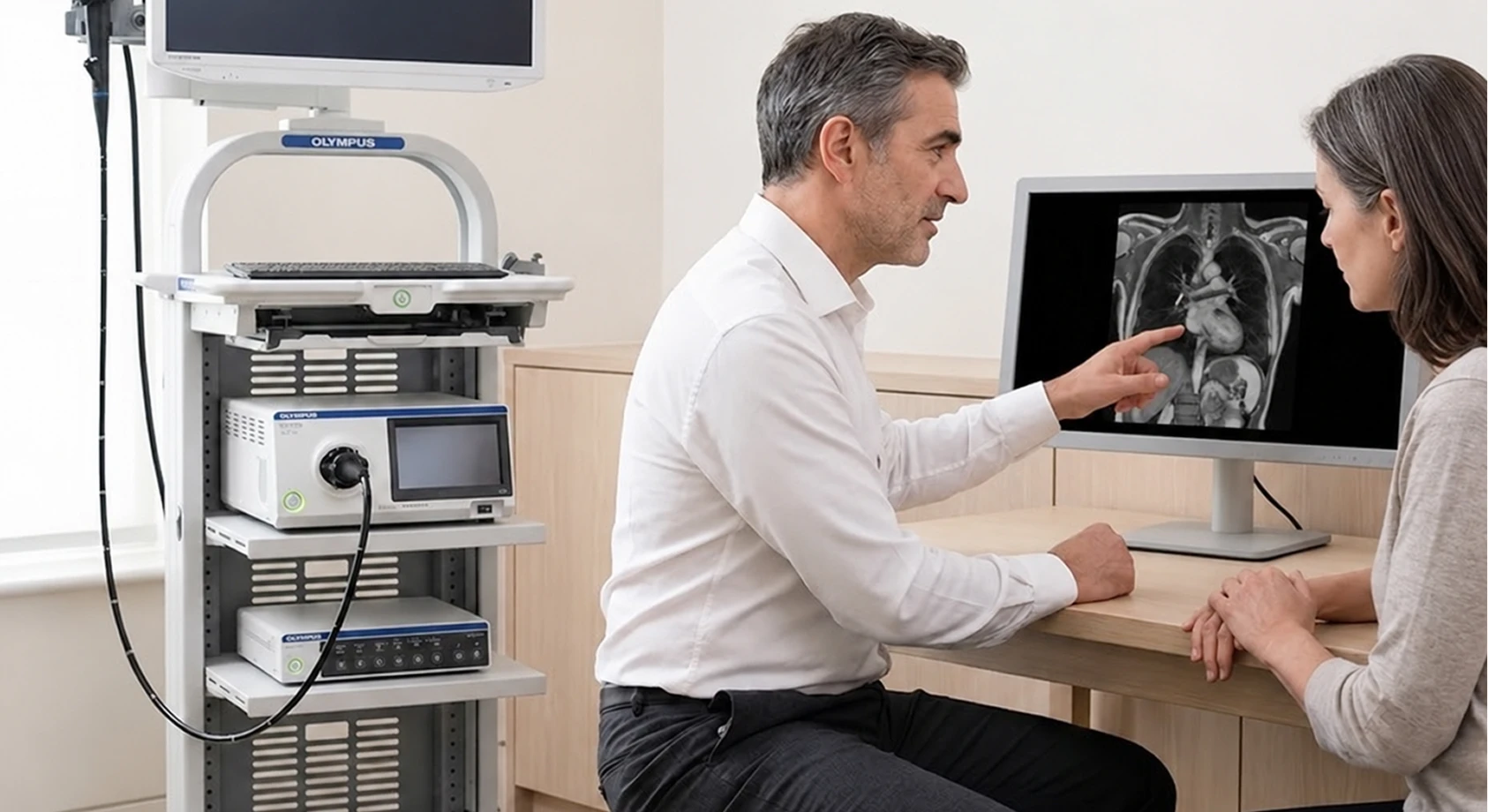
Some cancers are found more reliably by direct visualisation or by analysis of blood or tissue than by imaging, and the department uses each where it outperforms the alternative. Colonoscopy and gastroscopy are performed on the Olympus EVIS X1, whose artificial intelligence flags polyps in real time, with faecal immunochemical testing used too. Prostate-specific antigen is measured with free PSA and density and interpreted alongside imaging rather than as a number. More generally, tumour markers are used where indicated and read as part of a picture and circulating tumour DNA analysis offers a further route to detection.
Skin
Skin cancer is among the most curable cancers when it is found early. The department examines the whole skin surface, assesses every lesion of interest by dermoscopy, and records the entire skin by total body photography on the FotoFinder ATBM master.
Hereditary risk
Inherited predisposition changes both the intensity of surveillance and the options available to reduce risk, and it concerns a family rather than a person. The department establishes it through testing for BRCA1 and BRCA2, Lynch syndrome and extended predisposition panels, with counselling before and after. Risk-reducing options are discussed plainly where they apply, and cascade testing is offered to relatives who share the risk, arranged and counselled through the Institute with Genomic and precision medicine.
Surveillance after treatment
For patients treated for cancer elsewhere, the department provides follow-up coordinated with the treating team, so that surveillance continues to the same standard once active treatment ends. Surveillance imaging and laboratory monitoring are undertaken at the intervals the cancer warrants. The long-term effects of treatment on the heart, the bones, the hormones and the metabolism are assessed and managed, and rehabilitation and recovery are planned.
Research collaboration
Detection improves only as fast as the evidence behind it, and no single clinic sees enough patients to move that evidence forward alone. The Institute contributes anonymised data, under formal agreement, to partner academic and clinical institutions, and takes part in multi-centre studies of early detection and hereditary risk. Research consent is sought and recorded separately from consent to treatment and is never a condition of care; findings are reviewed for clinical relevance before anything is published; and the governance of the whole is aligned with United Kingdom data protection law and research ethics standards.

